A 40-year-old female patient presented with a failing amalgam restoration with recurrent caries and with fracture on the distal marginal ridge on the upper left first molar (tooth No. 14) (Figure 1). The preoperative bite-wing was taken to make sure that there were no other interproximal caries (Figure 2). All the photos were taken with a CS 1200 intraoral camera (Carestream Dental). After applying a local buccal infiltration using 2% lidocaine with 1:100,000 epinephrine, a clamp No. 24 (Hu-Friedy) was placed on the second molar and a rubber dam was inserted.
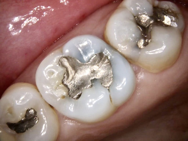
Figure 1. Preoperative photo of the tooth with failing amalgam restoration.
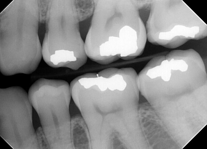
Figure 2. Bite-wing x-ray of the tooth.
The existing, old amalgam restoration was removed using a 806.31.009 inverted cone diamond bur (Brasseler USA). The recurrent caries and amalgam stains were visible (Figure 3). All the soft caries were removed using carbide round burs (sizes RA 2,4,6) with a Midwest Shorty slow-speed handpiece (Dentsply Sirona) (Figure 4). To make a clean and neat cavity preparation, all the superficial caries and stains were removed conservatively with a 6801.31.009 small round diamond bur (Brasseler USA). Then an intraoral photo was taken to confirm that no caries were left behind (Figure 5).
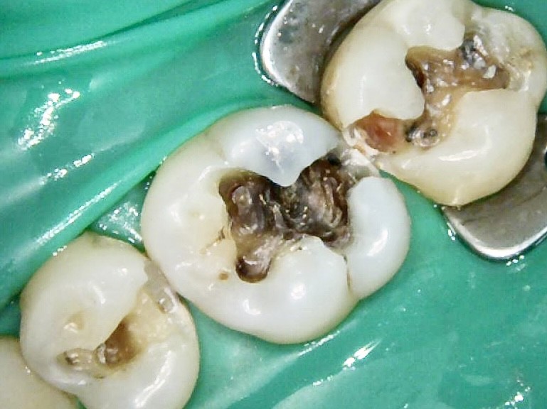
Figure 3. The tooth with recurrent caries.

Figure 4. Soft caries being removed.
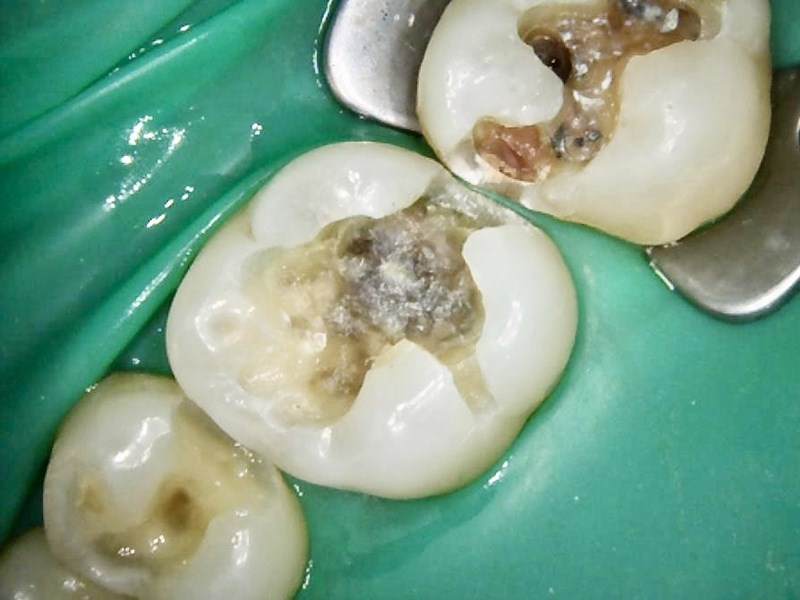
Figure 5. The tooth after all the caries were removed.
After a clean and neat cavity preparation, the tooth was dried well and TheraCal LC (BISCO), a light-cured, resin-modified calcium silicate material, was placed on the deep cavity surfaces and light cured for 20 seconds using the FlashMax P3 curing light (CMS Dental) to protect the pulp (Figure 6). Because TheraCal LC is opaque, it was also used to mask the dark amalgam stains that could show through the translucency of the composite. Due to the wide depth of the cavity preparation, TheraBase (BISCO) was also placed (Figure 7) and light cured for 20 seconds. TheraBase is a dual-cured base/liner that chemically bonds to the tooth structure and releases calcium and fluoride ions.
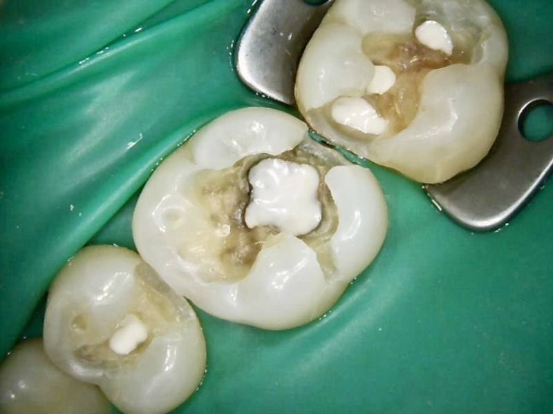
Figure 6. After the placement of TheraCal LC (BISCO).
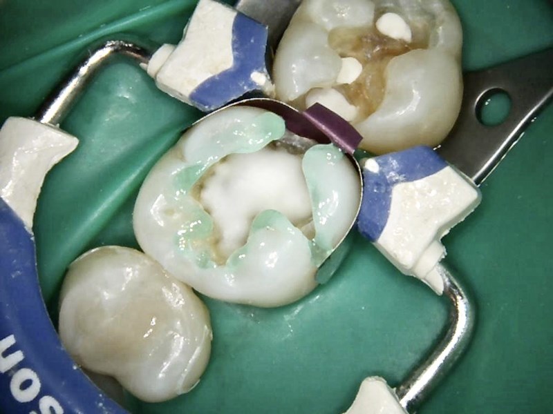
Figure 7. After TheraBase (BISCO) was placed.
After the placement of the liner and base, a blue ring and sectional matrix (Composi-Tight 3D Fusion Sectional Matrix System [Garrison Dental Solutions]) were placed (Figure 8). Then selective enamel etching was done using 37% phosphoric acid (Figure 9). The area was rinsed and dried, and All-Bond Universal adhesive (BISCO) was applied and light cured.
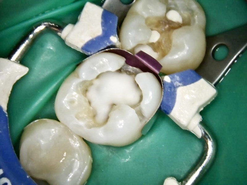
Figure 8. After the placement of a sectional matrix.
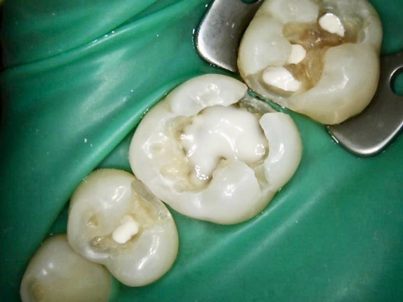
Figure 9. Selective enamel etching. A Restorative Approach Using Calcium-Releasing Materials
Estelite Sigma Quick composite (Tokuyama Dental America) was placed into the gingival box first in small increments using a condenser and plastic instrument (Hu-Friedy). Before light curing the final layer, some occlusal anatomy was created using several hand instruments.
The occlusal adjustments and more detailed anatomy were created using flame-shape diamond bur 274.31.016 from Brasseler USA. Finishing and polishing were completed using a large, coarse Sof-Lex disc (3M), as well as a No. 12 blade and a White Arkansas Stone (Dedeco). The final restoration looks natural and blends nicely to the tooth (Figure 10).
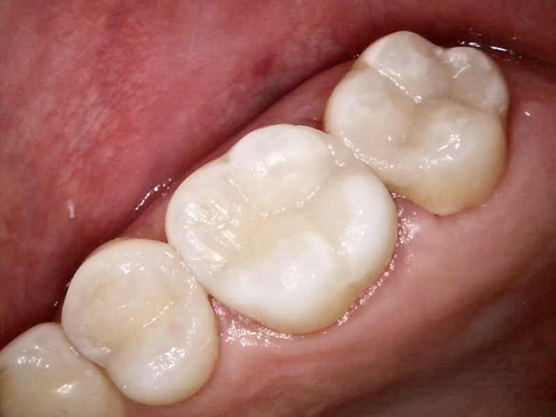
Figure 10. Final restoration.
Written by Dr James Chae